 Miscellaneous
Miscellaneous
Miscellaneous  Our World
 10 Green Practices That Actually Make a Difference
 Humans
 Ten Historic Men Who Deserve Way More Credit Than They Got
 Movies and TV
 The 10 Most Heartwarming Moments in Pixar Films
 Travel
 Top 10 Religious Architectural Marvels
 Creepy
 10 Haunted Places in Alabama
 History
 Top 10 Tragic Facts about England’s 9 Days Queen
 Food
Food 10 Weird Foods Inspired by Your Favorite Movies
 Religion
 10 Mind-Blowing Claims and Messages Hidden in the Bible Code
 Facts
Facts 10 Things You Never Knew about the History of Gambling
Miscellaneous Ten Groundbreaking Tattoos with Fascinating Backstories
Our World 10 Green Practices That Actually Make a Difference
Humans Ten Historic Men Who Deserve Way More Credit Than They Got
Who's Behind Listverse?

Jamie Frater
Head Editor
Jamie founded Listverse due to an insatiable desire to share fascinating, obscure, and bizarre facts. He has been a guest speaker on numerous national radio and television stations and is a five time published author.
More About Us
Movies and TV The 10 Most Heartwarming Moments in Pixar Films
Travel Top 10 Religious Architectural Marvels
Creepy 10 Haunted Places in Alabama
History Top 10 Tragic Facts about England’s 9 Days Queen
Food 10 Weird Foods Inspired by Your Favorite Movies
Religion 10 Mind-Blowing Claims and Messages Hidden in the Bible Code
Facts 10 Things You Never Knew about the History of Gambling
10 Fascinating Facts About The Human Skeleton
The skeleton might seem less dynamic than many of the human body’s other organ systems. Yet, the skeleton has many remarkable physical attributes that help it to support the human body as well as some truly remarkable biochemical attributes that regulate the functioning of the body. Here, we take the skeleton out of the closet for closer examination.
10 The Skeleton Influences Sugar Metabolism
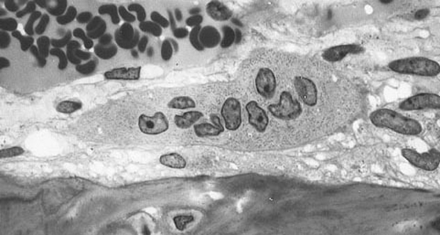
Photo credit: Robert M. Hunt
The skeleton is actually part of the endocrine system and a regulator of sugar metabolism, and it influences the manner in which certain fats are metabolized in the body. In 2007, researchers at the Columbia University Medical Center determined that human bone cells regulate blood sugar levels and fat deposition through secretion of the hormone osteocalcin. Osteocalcin increases insulin secretion, but without the decrease in insulin sensitivity that is normally seen in association with increased insulin secretion. Furthermore, osteocalcin boosts the number of insulin-producing pancreatic B-cells. The chemical also mitigates the storage of fat. It has become clear that the skeleton is an important metabolic regulator with a strong influence on how our bodies regulate the metabolism of sugar as well as weight gain and loss.
As a result, this function of our skeletal system plays a significant role in addressing the problem of type 2 diabetes, since osteocalcin levels are low in those afflicted. With that role comes the potential for mitigation of diabetes through medical intervention. According to Gerard Karsenty, chair of the department of Genetics and Development at Columbia University Medical Center, “The discovery that our bones are responsible for regulating blood sugar in ways that were not known before completely changes our understanding of the function of the skeleton and uncovers a crucial aspect of energy metabolism. These results uncover an important aspect of endocrinology that was unappreciated until now.”
9 Automatic Bone Replacement
Bone Remodeling and Modeling
Developing well before birth and growing in size over the years, the human skeleton might be viewed by a layperson as analogous to a steel building under construction. Gradually gaining in size, strength, and mineral content, the human skeleton is not simply built once. In fact, it changes over one’s lifetime—the most significant change being the gradual replacement of bone on a continual basis, which leads to replacement of the entire structure of each bone every 10 years on average.
In the younger years of life, a formation process known as modeling creates the opportunity for bone to form while old bone material is removed from a second site inside that particular bone, enabling proper bone growth. Remodeling, however, takes place over the course of one’s life, becoming the primary means of bone structure change by one’s early twenties. Through remodeling, most of the adult skeleton is fully replaced approximately every 10 years. The complex processes associated with modeling and remodeling, known as bone metabolism, involve five stages of biochemical activity, including digestion of bone material and subsequent rebuilding of new bone structures.
8 Gorham’s Disease
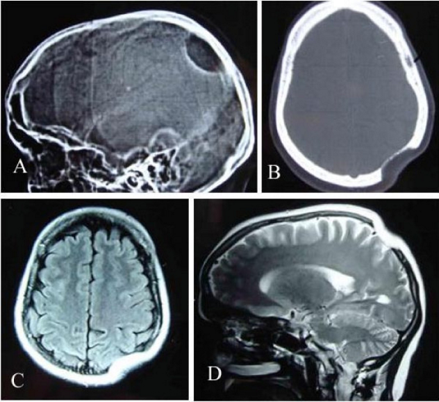
Photo credit: Parihar V., Yadav Y.R., Sharma D.
A system as strong, complex, and biologically active as the skeleton also has its weak points. Like the rest of the body, the skeleton can succumb to a variety of medical challenges, some common, some rare and unusual in nature. The existence of Gorham’s disease is an example of just how insidious bone-related disease and dysfunction can be. Defined by bone loss, or osteolysis, in specific areas of the body, Gorham’s disease–associated bone loss may occur anywhere in the human skeleton.
However, it occurs with the greatest frequency in the skull, shoulder, rib, jaw, spine, and pelvic bones, where it causes wasting of the bone. The disease can also affect soft tissue and nearby bone structures, leading to further damage and weakening. According to the National Organization for Rare Disorders, Gorham’s disease, eerily known by the alternate name “vanishing bone disease,” may even lead to death if the spine is significantly affected or lung function compromised.
The root cause of this rare disorder is a mystery. There is no single way to address Gorham’s disease, but a variety of approaches have been tried in various situations, ranging from surgery on affected areas to use of bone resorption- or lymphatic vessel formation–inhibiting medications.
7 The Incredible Hyoid Bone
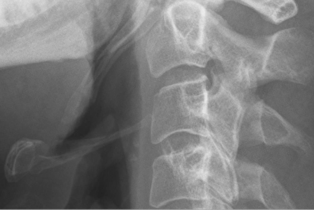
Photo credit: Hellerhoff
The hyoid bone is considered anatomically separate from the larynx. It is unique among all the bones in the body, as it’s anatomically isolated from every other bone in the skeleton. Nested among cartilage and supporting the larynx, the hyoid bone is remarkable not only for its physical structure and skeletal isolation, but also for its foundational and pivotal impact on human evolution. Providing the anchor for muscles associated with the tongue and mouth floor muscles, the hyoid is complex in structure, with a center and projecting horns that give it a U-shaped appearance. The hyoid bone consists of three primary parts—the body of the hyoid, greater cornua, and the lesser cornua .
Through the development of the highly complex hyoid bone, adapted to work in unison with the rest of the larynx, human speech had the opportunity to develop to a much greater degree than in other mammal species. The complex structure of the hyoid bone and larynx working in together in a finely orchestrated manner supports the articulation of complex sounds in humans.
A development supported by the hyoid bone takes place with age—the physical drop of the larynx in infants, which creates a drop in vocal pitch and also makes speech possible. During puberty, a further drop of the larynx and vocal pitch takes place in young males. Interestingly, these developments parallel evolutionary history, where the drop in the larynx supported the development of human speech.
6 The Incredible Resilience Of The Human Jaw
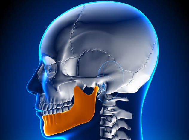
The hardest bone in the human body could be a range of things upon initial thought. One might imagine that it’s the femur, due to its resistance to breakage. The hard, knobby heel might come to mind, or perhaps the elbows. The strongest bone in the body (and the largest single bone in the skull) is actually the mandible, aka the lower jaw. The relatively massive bone is the only mobile skull bone, able to hold the teeth and move an enormous amount during one’s lifetime while withstanding repeated and significant levels of stress.
Meeting the rest of the head at almost at right angles, the hardness of this bone allows it to be streamlined and precisely fitted to efficiently accomplish its tasks while being small enough to remain in scale with the rest of the head. The hardness of this bone exceeds that of every other bone in the human body and is indeed remarkable, showing the evolutionary power of necessity in varying the hardness of human bones exactly in relation to their jobs. While broken jaws do occur, they are far less likely than the slim form of the jawbone suggests, thanks to this remarkable hardness.
5 Of Bones And Bloodstreams
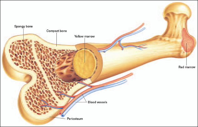
Photo via the Foundation for Biomedical Research
One might put the bones and the blood cells together last when thinking of closely associated body system elements. However, the truth of the matter is that skeletal red and white blood cell production underpins our survival as human beings. This is because the marrow tucked inside our some of our bones plays a vital role in the formation of our bloodstream components, forming red and white blood cells and platelets. In the very young, the need for blood cell production is high, with the majority of the bone marrow consists of red, or hematopoietic marrow, distributed all throughout the body. In infants, red bone marrow may even be found in the fingers. With age, more and more of it is converted to the yellow type.
Located in adults across a more limited extent of bone structure, red marrow occurs in the hip bones, sternum, ribs, vertebrae, shoulders, and skull bones, plus the spongy material in the femurs and humeri. On average 2.6 kilograms (5.7 lb) of bone marrow exists in the body. As adults mature, much of the red bone marrow gradually gives way to yellow bone marrow, which produces fat.
How do blood cells produced in the bones end up in the circulatory system, one might ask? The answer is intricate, logical, and thought-provoking. The vascular bone marrow is filled with capillaries and blood vessels. Once formed, cells migrate through sinusoid cavities into the main components of the bloodstream.
4 The Pelvis, Hormones, And Human Birth
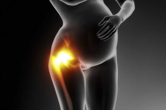
In order to accommodate the challenge of birthing a human baby, particularly with its exceptionally large cranium, the female human body has developed some remarkable adaptations. One of the most interesting skeletal adaptations involves hormonally influenced changes affecting the laxity of the pelvis joints, thanks to a hormone aptly named relaxin.
Relaxin, produced in the human reproductive system, has a significant effect in females on the cervix but also on the smooth muscles, ligaments, and joints of the pelvis. Pelvic joints become more stretchable thanks to relaxin’s general loosening effect, aiding and abetting delivery of the baby. However, it has been suggested that this stretching and loosening effect could make those affected more unsteady on their feet while pregnant.
In an article in a Scanadinavian journal of medicine and science, relaxin is described as “mammalian 6-kDa heterodimeric polypeptide hormone” and “a member of the insulin-like superfamily.” The article points to a range of fascinating areas of study, such as animal research–based findings and human health considerations relating to the interaction between relaxin and the musculoskeletal system. Studies cited found a fourfold increase in the rate of anterior cruciate ligament (ACL) injuries in elite female athletes with relaxin concentrations above 6.0pg/mL. Research also found an association between the occurrence of ACL injuries and the menstrual cycle, with injuries being more frequent during the ovulatory phase.
Other human studies suggested a link between pelvic instability and weakness in other joints with elevated relaxin levels, while animal studies have repeatedly shown such correlations to be strong in other species. Problems may have the potential to arise when relaxin is used as a therapy in certain cases.
3 Melorheostosis
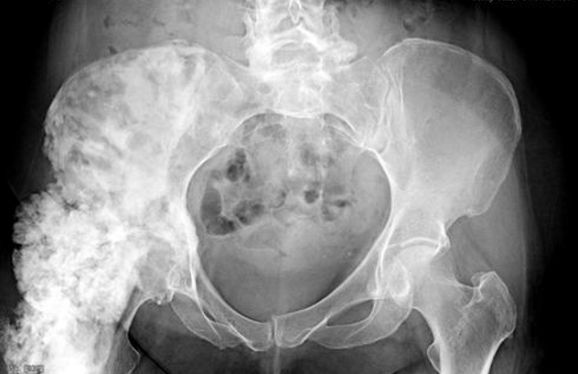
Photo via USA Today
A rare disease that stands out by presenting one of the most disturbing and unusual fates that the human skeleton can face, melorheostosis is a mesenchymal dysplasia that affects just one in a million people and presents mysteries and challenges that continue to confound medical science. Melorheostosis results in the growth of exceptionally hard new bone material on top of existing bone in an erratic manner that has a flowing appearance. The appearance of the invasive growths can resemble candle wax when viewed in an X-ray image.
The disease can emerge as a result of genetic predisposition, but environmental factors may exacerbate or mitigate occurrence potential. Oddly, a case was described involving identical twins, one of whom had the condition and one of whom who did not. Additionally, the disease has been described as weird and unusual, according to an orthopedic oncologist from the Mayo Clinic, with a “broad spectrum of symptoms” found in patients unlucky enough to have the condition. According to the Melorheostosis Association, effects can include disabiling pain, soft tissue symptoms, deformity, and severe functional limitation of affected body areas.
2 Skull Ridges And Brain Injury
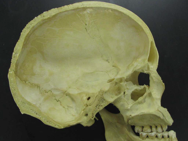
Photo credit: Tim McCormack
Our heads can work against us, literally when it comes to traumatic brain injury suffered through impacts with the skull. Our strong skulls may generally protect us from hard knocks and prevent actual impact to the brain from an outside object. However, the brain is not securely anchored in the skull, and room between the brain and the skull allows for movement. If the human head moves quickly, the brain is moving inside the skull. In the case of a sudden stop, the brain will keep moving due to inertia until it crashes into the interior of the skull, while a blow to the head will create shock waves that also lead to brain movement, causing the brain to hit the skull.
Remote damage can also occur due to shock wave transmission, and that is where the anatomy of the skull can exacerbate brain injury. Bony ridges lining the base of the skull can injure the surface of the brain upon impact, leading to tears, lacerations, and related injury through collision with the sharp surfaces. The movement of the brain and the forces involved can also stretch nerve axons and tear blood vessels. In fact, most traumatic brain injuries occur without the skull being penetrated. Injuries can occur to one side of the brain or the opposite side as well if the brain moves back and forth. Injuries where the impact results in a collision with the side of the head may also lead to brain damage through bruising.
1 Smoking Bones

Smoking is widely recognized as being bad for your health, and smoking is bad for your bones, too. Osteoporosis is one of the leading causes of bone degeneration, and affected individuals are at a greater risk bone fractures. Research shows that smoking predisposes individuals to increased rates of osteoporosis through malnutrition of the bones. Smoking deprives your bones of calcium by impeding the body’s use of Vitamin D, which would otherwise assist your body in transferring calcium into the bones. The result? Fragile bones. Smoking also poisons your osteoblasts, or bone-forming cells.
Furthermore, smoking cuts into estrogen production in both males and females. Estrogen increases the ability of the bones to retain calcium. Smoking while bones are being built reduces their ultimate mass, while smoking after the age of 30 increases the rate of bone loss up to twofold. The hip, spine, and wrist bones are at greatest risk. Osteoporosis rates may be 2.5 times as great as in nonsmokers. Female smokers may have 15 to 30 percent reductions in bone mineral levels, while males may see losses of 10 to 20 percent. According to the World Health Organization, studies indicate that one in eight hip fracture cases arise from smoking.
Christopher M. Stephens, Msc., is an ecologist and conservation planning consultant from British Columbia, Canada, with a passion for discovering the most startling secrets of our natural world. He is the birding tour leader for Pacific Rainforest Adventure Tours, offering international visitors the opportunity to see remarkable Canadian birds up close and accepts custom bookings throughout the year.
http://rainforestnaturehikes.com/?p=20
More Great Lists
 10 Fascinating Facts About Human Evolution
 Top 10 Fascinating Feats Of The Human Voice
 10 Facts About Human Cannibalism From Modern Science
 10 Unnerving Facts About Human Violence From Modern Science
 10 Horrifying Facts About The Human Form Of Mad Cow Disease
 10 Remarkable Facts About Animals Put In Human Perspective
 Top 10 Fascinating Facts About Magnetic Fields
 10 Fascinating Facts About The American Flag
 10 Fascinating Facts About The Bugs Living In Your Guts
fact checked by
Jamie Frater
More Great Lists