 Misconceptions
Misconceptions
Misconceptions  Creepy
 10 Prestigious Universities with Famous Ghost Stories
 History
 10 Daring Military Operations That Ended in Disaster
 Movies and TV
 10 Amazing Lost Films That Were Found Again
 History
 10 Wild Stories About America’s Most Fascinating Founding Father
 Our World
 10 Astonishingly Valuable Things Their Owners Simply Walked Away From
 Humans
 10 Disturbing Communities from the Dark Corners of the Internet
 Movies and TV
 10 Hollywood Style Choices That Backfired
 Weird Stuff
 10 Ancient Chores That Would Horrify Modern Health Inspectors
 Politics
 10 Strange High-Tech Tools Shaping Modern Politics
Misconceptions 10 Common Misconceptions About Famous Scientists
Creepy 10 Prestigious Universities with Famous Ghost Stories
History 10 Daring Military Operations That Ended in Disaster
Who's Behind Listverse?

Jamie Frater
Head Editor
Jamie founded Listverse due to an insatiable desire to share fascinating, obscure, and bizarre facts. He has been a guest speaker on numerous national radio and television stations and is a five time published author.
More About Us
Movies and TV 10 Amazing Lost Films That Were Found Again
History 10 Wild Stories About America’s Most Fascinating Founding Father
Our World 10 Astonishingly Valuable Things Their Owners Simply Walked Away From
Humans 10 Disturbing Communities from the Dark Corners of the Internet
Movies and TV 10 Hollywood Style Choices That Backfired
Weird Stuff 10 Ancient Chores That Would Horrify Modern Health Inspectors
Politics 10 Strange High-Tech Tools Shaping Modern Politics
10 Freakishly Large Single-Celled Organisms
In general, the term “single-celled organism” is synonymous with being microscopic, and not without good reason. The vast majority of unicellular organisms never grow more than one tenth of a millimeter long. Their size is limited by several factors: It’s harder for large cells to retain structural integrity; transporting food and waste from one part of the cell to another becomes difficult. In many cases, growing bigger just wouldn’t provide enough of an evolutionary benefit to justify putting all the energy into extra growth. These and other factors help keep microbes just that—microscopic. Yet in a realm as old, vast, and diverse as the microbial world, there are bound to be exceptions. This list is devoted to some of those single-celled “microbes” that are anything but microscopic.
10 Stentor
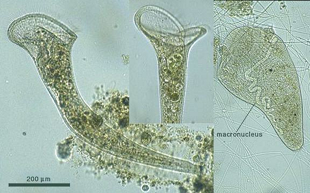
Photo credit: Protist Image Database
Growing up to 2 milimeters long, the trumpet-shaped freshwater protozoa of the genus Stentor are easily visible to the naked eye and well-known among microbe enthusiasts for their size. 2 millimeters might not sound impressive, but remember that this makes Stentor larger than many multicellular invertebrates. Among unicellular organisms, it is an absolute colossus.
One of the factors that enables Stentor to get so big is its internal anatomy. Unlike regular cells, Stentors (like most of the entries on this list) have more than one nucleus, the part of a cell that houses its DNA and acts as its control center. Having multiple nuclei seems to make it easier for bigger cells to properly manage their relatively large cellular bodies. Specifically in Stentor’s case, it has numerous small micronuclei that control reproduction and a single, giant, string-like macronucleus that manages its regular functions.
Stentors are what biologists call a ciliate; they’re covered in fine, hair-like structures called cilia. Stentors and other ciliates use these to swim, beating them in unison to propel themselves, but that isn’t all cilia can do. While Stentors gain some nutrients from symbiotic algae that often lives inside them, they are primarily filter feeders. To catch food, Stentors anchor themselves to floating debris or sediment, unfold their trumpet-like “mouth,” and use a ring of modified feeding cilia to create a current that sucks in bacteria, smaller protists, and the occasional unlucky water flea.
In other words, not only is the unicellular Stentor bigger than several multicellular animals, but it sometimes eats them.
9 Spirostomum
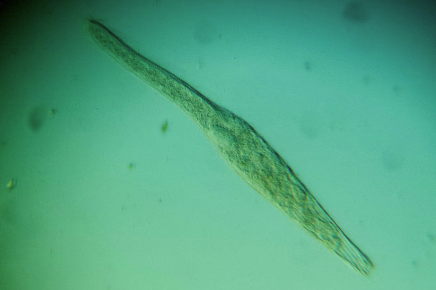
Photo credit: Picturepest
With the largest species growing up to 4 millimeters long, members of the worm-like Spirostomum genus dwarf their Stentor relatives. Found both in fresh and saltwater, it is often mistaken for a small worm. When viewed under a microscope, though, it becomes clear that it is in fact a single, really long cell.
Despite its length, Spirostomum is also notable in the microbial world for its incredible shrinking ability. When it is disturbed, it can shrink down to a quarter of its original size in less than a hundredth of a second. This is the fastest-known contraction of any cell.
Like Stentor, Spirostomum is a ciliate. The cilia are arranged in a spiral formation and both propel it forward and sweep bacteria into its small “mouth” along the side of its body. Also like Stentor, Spirostomum has one large macronucleus and multiple smaller micronuclei. This setup is largely unique to ciliates.
They do differ from Stentor in terms of prey, though. While Stentors are big game hunters that can take down small multicellular life, Spirostomum mostly sticks to bacteria.
8 Chaos Carolinensis
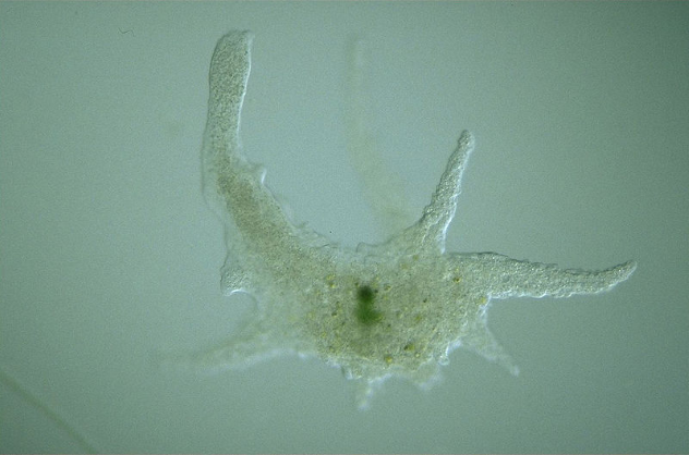
Photo credit: Dr. Tsukii Yuuji
Picture an amoeba. Now enlarge it to the size of a sesame seed. You have Chaos carolinensis. While their exact dimensions change with their shape, the largest individuals can stretch to 5 millimeters in length. It is so big that putting a cover slip on it under a microscope can harm it.
Despite its large size, C. carolinensis behaves much the same way as a smaller amoeba would. It moves around using temporary gelatinous protrusions called pseudopods (Latin for “false foot”). It also uses these to feed. When it encounters prey, C. carolinensis literally engulfs it with its pseudopods and absorbs the prey in an internal, temporary cavity called a vacuole. There, the prey is digested alive, and the remains will eventually be expelled from the cell as waste. C. carolinensis feeds on other microbes as well as small invertebrates like water fleas or rotifers. It will continue feeding until it’s ready to reproduce.
Like Stentor and Spirostomum, C. carolinensis has multiple nuclei, although they aren’t organized or specialized like in the other two. A single nucleus simply would not be able to control a cell this big. In fact, depending on its size, C. carolinensis can have up to 1,000 nuclei.
Chaos carolinensis was subject to a decades-long naming controversy after its discovery, as scientists argued over how to classify it. For this reason, older sources referred to it by a variety of names, including Pelomyxa carolinensis and Chaos chaos. To avoid confusion, some writers simply introduced the protist as “the giant amoeba.”
7 Gromia Sphaerica
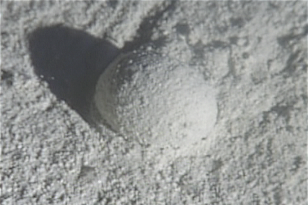
Photo credit: Mikhail Matz
When researchers from the University of Texas dove to the seafloor off the Bahamas, they were baffled to find dozens of odd, grape-sized balls that, despite seeming motionless, had clearly left trails in the sand. Initial guesses ranged from a strange new type of snail to oddly-shaped fecal matter. However, upon closer examination, the truth turned out to be even weirder. The balls were actually giant, 3-centimeter (1.2 in) wide spherical protists that were rolling across the seabed at a near glacial pace.
Gromia sphaerica, or the Bahamian Gromia, is what biologists call a testate amoeba. In other words, it is an amoeba-like creature that encases itself in a soft, porous shell called a test. By continuously sending out its thin pseudopods through holes in the test and grabbing onto the sea floor, the cell is able to slowly roll itself along the bottom, feeding on organic matter in the sediment as it goes.
The discovery of this gentle giant of a protist had dramatic implications for scientists’ understanding of the evolutionary timeline. The earliest conclusive evidence for multicellular life dates back to 580 million years ago, but the discovery of fossilized tracks dating as far back as 1.8 billion years ago has led some scientists to push the starting date back to much earlier. Surely, they argued, no microbe could have produced them. Yet it turns out that those fossilized tracks bear a strong resemblance to those of G. sphaerica, meaning that its ancestors may have produced them. Thus, the earlier starting date for multicellular life seems much less likely.
Unfortunately, not much else is known about these rolling blobs of cytoplasm due to the difficulty of taking live samples. Despite having a kind of shell, they are squishy and fragile by our standards. Researchers have described them as softer than a grape.
6 Sailor’s Eyeball
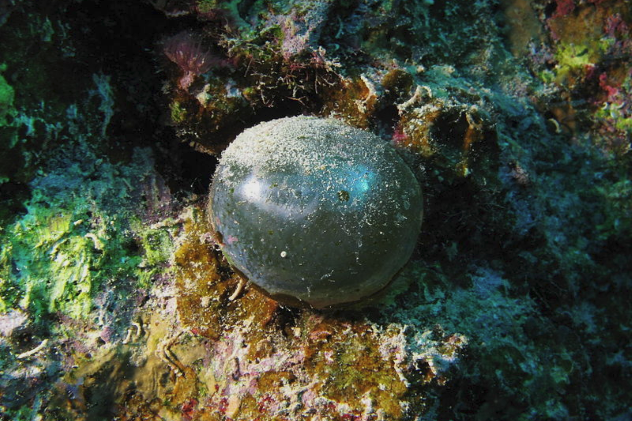
Photo credit: Alexander Vasenin
So far, all of the entries on this list have been “animal-like” protozoa, but in fact, there could be an entire list devoted to giant unicellular algae. Also known as bubble algae, Sailor’s Eyeball (Valonia ventricosa) easily grows to 4 centimeters (1.6 in) in diameter or more. Found in shallow tropical waters across the world, this marble-like protist is usually solitary but is sometimes found living in small clumps. Younger individuals have a beautiful translucent green color, but older ones are often encrusted with smaller types of algae and animals. In other words, Sailor’s Eyeball is so large that some multicellular life-forms actually live on it.
Although some admire it for its peculiar biology and exotic, gemstone-like appearance, Sailor’s Eyeball is best known as a despised pest for aquarium enthusiasts. Often accidentally introduced into tanks when owners bring in “live rocks” taken from the ocean, the algae goes on to overrun the tank, and killing or removing it is surprisingly difficult. Popping them is no use, either, since that’s actually how they reproduce.
5 Spiculosiphon Oceana
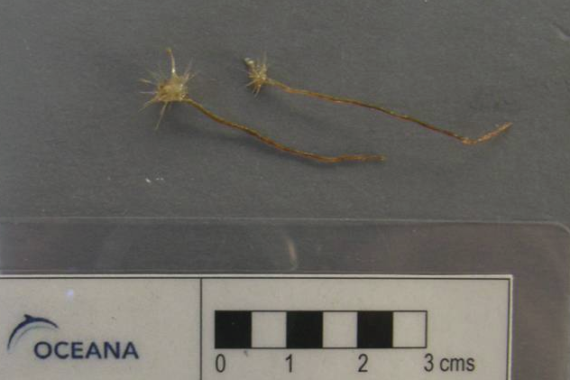
Photo credit: Silvia Garcia
With a maximum length of 5 centimeters (2 in), this strange aquatic protozoan has amazed scientists from the moment they first documented it. When divers first found it in 2013 in an underwater cave off the coast of Spain, they initially mistook it for a carnivorous sponge. (Yes, such sponges do exist.) However, this wasn’t the case.
Spiculosiphon oceana belongs to a type of test-building amoeba called Foraminifera, but being a “testate amoeba” is about the only thing that it has in common with its not-so close relative Gromia sphaerica. Unlike the rolling, detritus-eating sea-grape, this one is fixed in place and is a filter feeder. To catch food, S. oceana simply extends its long, tentacle-like pseudopods through the pores in its test and lets them float in the water, trapping and digesting any plankton that gets ensnared. In this manner, S. oceana’s feeding strategy is remarkably similar to many marine invertebrates, including carnivorous sponges.
For the very accomplishment of being a 5-centimeter-long unicellular organism, scientists named S. oceana one of the top 10 new species discovered in 2013.
4 Acetabularia
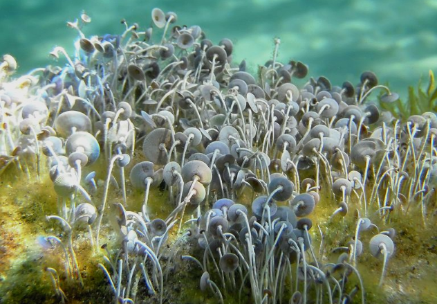
Photo credit: Tigerente
Also known as Mermaid’s Wineglass, Acetabularia is a unique genus of mushroom-shaped algae that grows up to 10 centimeters (4 in) in height. Found primarily living in clusters in shallow, rocky waters, it lives in subtropical waters around the world and they sometimes carpet large spots of seabed with their light green caps.
Acetabularia significantly differs from the other entries on this list in terms of its internal composition. As discussed earlier, large unicellular organisms usually have more than one nucleus, and the number generally increases with size. Yet despite dwarfing all the previous entries, Acetabularia spends most of its life with only a single, giant nucleus located at the base of its “stem.” The only exception is when it is about to reproduce. At this point, the nucleus undergoes multiple rounds of division, and the daughter nuclei travel up to the cell’s top frond. There, they bud off into numerous spore-like reproductive cysts, ready to spread and give rise to new Acetabularia.
The cell’s large size combined with its reliance of a single nucleus gave it a key role in the advancement of cellular biology. In a set of experiments during the 1930s and 1940s, German scientist Joachim Hammerling (whose work was funded by the Nazis) proved that the nucleus was the control center of a cell by grafting together the caps and nuclei of two species of Acetabularia. He found that the cell would take on the characteristics of whichever species its nucleus came from.
3 Syringammina Fragilissima
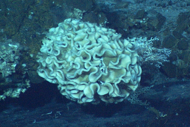
Photo credit: NOAA
The largest member of the Xenophyophore class (example pictured above), which already is known for producing unicellular giants, this giant amoeboid creature dwells at the bottom of the ocean and can grow up to 20 centimeters (8 in) in diameter. Like most of its relatives, the cell does not produce its own test but instead constructs it from the remains of smaller microorganisms and sponges. It glues these together with a slimy excretion to form a complex network of delicate tubes, which serve as the Amoeba’s home.
Unfortunately, we still know very little about Syringammina fragilissima. Scientists suspect that it feeds on bacteria, but they don’t know how it does so. Guesses range from filter feeding to farming them inside its shell. Scientists aren’t even sure how S. fragilissima reproduces. Part of the issue is the creature’s deep-sea habitat, but it also has to do with its extremely delicate nature. Its scientific name means “very fragile sand pipe.”
2 Plasmodial Slime Molds
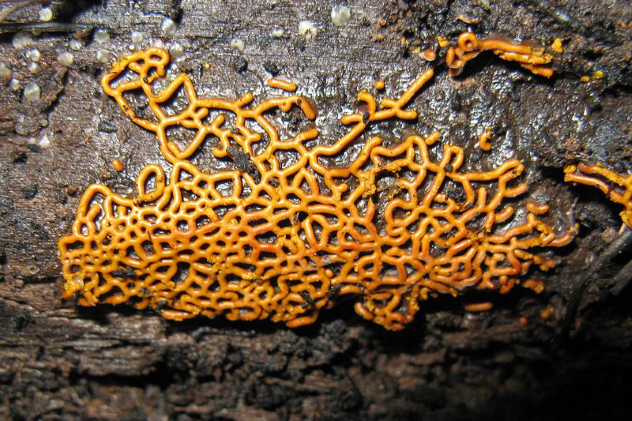
Photo credit: John Carl Jacobs
Originally classified as a type of fungus, plasmodial slime molds, also known as Myxomycetes, are an unusual category of unicellular life that blur the boundary between an individual organism and a group of them. Like all slime molds, they start life out as tiny, amoeba-like microbes that live in the dirt much like a regular single-celled organism, munching on bacteria. Under certain conditions though, something changes. The individual cells congregate together and begin to combine until they have merged into one colossal blob. Although most slime molds remain small by our standards even in this form, a few can grow to more than 1 meter (3 ft) in diameter, if not more.
Now living as a single organism, the slime mold will start to crawl across the ground at a glacial pace, consuming whatever food or unfortunate bacteria falls into its path. In essence, it acts like a giant amoeba and is capable of navigating around obstacles and sensing the best food sources from afar. This phase continues until it has eaten enough. At that point, the slimd mold will stop moving, produce fruiting bodies, and release spores to start the cycle anew.
But wait. If it originated from individual cells gathering together, then isn’t the slime mold not technically unicellular? Nope. Plasmodial slime molds truly are unicellular. Unlike the so-called “cellular slime molds,” in which the cells retain their distinct membranes, plasmodial slime mold cells fuse completely, dissolving the membranes separating one another and becoming a single, gargantuan cell with millions of nuclei.
1 Caulerpa Taxifolia (Aquarium Strain)
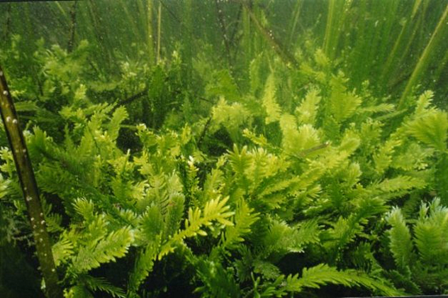
Photo credit: NOAA
Consisting of a long string of fern-like fronds, this type of unicellular algae is a giant even among its family of fellow macroscopic unicellular algae. In the Mediterranean, where it thrives best, it can reach a total length of nearly 3 meters (10 ft). Caulerpa taxifolia is so large, so structurally complex, and so multicellular-looking that some sources simply forget to mention that it is actually all one, unfathomably long cell with countless nuclei and other parts floating within.
C. taxifolia is not native to the Mediterranean, however, nor does it normally even come close to this size in its natural tropical habitat. Instead, the colossal Mediterranean variant is the result of human interference, somewhat like the Africanized killer bee. Attractive and easy to care for, C. taxifolia lends itself toward use in aquarium display tanks, and in the 1970s, a German aquarium acquired some of the algae in order to breed it for this exact purpose. Exposing their C. taxifolia to harsh chemicals and mutation-inducing UV light, the staff selectively cultivated it to be even hardier, faster growing, and most importantly, better able to grow in colder water. Finally, in 1980, they were satisfied, and in an act of generosity, they distributed the finished product to other aquariums across Europe.
Four years later, the inevitable happened. Some of the cold-water strain “escaped” from an aquarium in Monaco. Within years, it had overrun the Mediterranean. Compared to its natural ancestor, the mutant strain is bigger, grows faster and more aggressively, can survive pollution, and is capable of regenerating from fragments as small as 1 centimeter (2.1). It’s also toxic. Eradication efforts have failed, and the only question is how to keep it from spreading even farther.
Because of the ecological devastation it has brought, C. taxifolia earned the nickname “killer algae,” along with a place on the Global Invasive Species Specialist Group’s list of the 100 worst invasive species.
Regardless, there you go—a unicellular organism that’s bigger than you.
Robert Bellaskus is a bookish eccentric whose interests range from world history to deep-sea life.
More Great Lists


fact checked by
Jamie Frater
More Great Lists