 Weird Stuff
Weird Stuff
Weird Stuff  Mysteries
 10 Tragic Disappearances and Deaths in Joshua Tree National Park
 History
 10 Ways Childhood Really Sucked in the Old West
 Music
Music 10 Name Origins of Famous Bands from the 1990s
 Religion
 10 Biggest Turnarounds by the Catholic Church
 Weird Stuff
 10 Unbelievable Times Laws Had Unintended Consequences
 Humans
 Ten Historic Women Who Deserve Way More Credit Than They Got
 Movies and TV
 10 Films That Spawned Major Lawsuits
 History
 Ten Times Towns Were Wiped Off the Face of the Earth
 Creepy
 10 of the Most Disturbingly Haunted Public Houses in the UK
Weird Stuff 10 Niche Subcultures That Are More Popular Than You Might Think
Mysteries 10 Tragic Disappearances and Deaths in Joshua Tree National Park
History 10 Ways Childhood Really Sucked in the Old West
Who's Behind Listverse?

Jamie Frater
Head Editor
Jamie founded Listverse due to an insatiable desire to share fascinating, obscure, and bizarre facts. He has been a guest speaker on numerous national radio and television stations and is a five time published author.
More About Us
Music 10 Name Origins of Famous Bands from the 1990s
Religion 10 Biggest Turnarounds by the Catholic Church
Weird Stuff 10 Unbelievable Times Laws Had Unintended Consequences
Humans Ten Historic Women Who Deserve Way More Credit Than They Got
Movies and TV 10 Films That Spawned Major Lawsuits
History Ten Times Towns Were Wiped Off the Face of the Earth
Creepy 10 of the Most Disturbingly Haunted Public Houses in the UK
10 Body Parts That Are Secretly Awesome
Some body parts get all the attention, whether it’s the famous essentials like the heart, brain, and liver or the beauty of smiles or athletic musculature. However, there is a whole world of phenomenal body parts that deserve some more attention.
These unsung anatomical heroes might not be the most eye-catching, but they’re why you don’t walk into walls, choke every time you eat, or simply keel over dead while you’re reading this article, among other things. Here is a list of ten of the most underappreciated, interesting, and important parts of the human body.
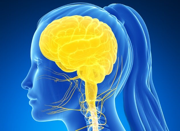
10 Vestibular System
Vestibulo-Ocular Reflex

Ever wondered how you know where your head is in space? How you don’t get dizzy every time you nod or tilt your head? Or why you can’t walk in a straight line after spinning in a circle for a long time?
The answer is the vestibular system (VS), a minuscule, complex setup comprised of three semicircular canals and two chambers in each inner ear. The VS sits behind your eardrum, just next to the cochlea. The semicircular canals are three round tubes filled with liquid, which lie in different planes, enabling sensation of movement in all directions. There are special areas called maculae (not to be confused with the maculae in the retinas) at the end of the tube loops which are covered with sensory hairs. On top of the hairs is a jelly-like substance with tiny weights in it called otoliths. When you move your head, the semicircular canals and maculae move, but the fluid and jelly lag behind. This lag bends the sensory hairs and sends a message to your brain about the direction your head is moving. When you stop moving (or accelerating) and keep your head in a particular spot, the effect of gravity on the weighted jelly tells your brain where you are in space.
So, what happens when we spin in a circle and get dizzy? Ask a friend to spin in a tight circle, either on their feet or in an office chair, for over 30 seconds and then suddenly stop and try to focus on a fixed point. They will feel dizzy and struggle to walk in a straight line, and if you look closely, you will see their eyes flicking from side to side (a phenomenon called nystagmus). This happens because your VS has stopped moving, but the fluid inside the loops has enough momentum to keep moving. This tells your brain you are spinning, but your eyes and cerebellum don’t agree, so you feel completely off-balance, and your vision is distorted.[1] You can also watch the medical student above try it.
9 Kneecaps
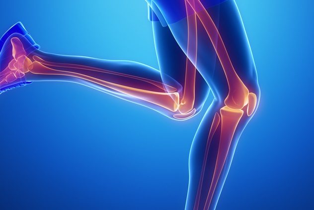
If you have ever fallen on your knees or had that sickening feeling of sliding a chair under a desk and colliding with an unfortunately placed table leg, you’ve probably been grateful for their protection. However, kneecaps are much more than built-in, rudimentary kneepads!
It’s all a matter of leverage. The main function of the kneecap, technically called the patella, is extension of the knee (straightening the leg). The kneecap is tethered to the shinbone (tibia) by a strong tendon, and the top of the kneecap is connected to a major muscle in the quadriceps group. Your “quads” are a group of four muscles, hence the name. The patella increases the effective force with which the knee can extend by 33 to 50 percent due to the increased leverage around the joint.[2]
8 Cerebrospinal Fluid
Amid all the flesh, blood, and guts in the human body is this beautiful, crystal-clear fluid. Cerebrospinal fluid (CSF) is produced in ventricles deep within the brain and circulates around the brain and spinal cord.
CSF has many functions, including protection, as it provides an area of shock-absorption for the brain when the skull is hit or shaken. It also works to provide nutrients and clear waste from the brain and spinal cord in a similar way to blood in other parts of the body. The CSF is produced and absorbed in an exquisite balance to maintain the correct pressure to surround and support the central nervous system (brain and spinal cord).
Doctors sample CSF by performing a procedure called a lumbar puncture—inserting a needle into the spinal cord and collecting some of the fluid.[3] It can be used to identify people who have an infection (such as meningitis), a bleed around the brain (hemorrhagic stroke), and other conditions.
7 Uterus

Most women are not particularly fond of their uterus, as it is often a source of pain or problems, but it deserves a prized place on this list.
The most obviously remarkable feature of the uterus is its ability to expand from approximately the size of a woman’s fist to fill most of the abdomen and some of the thorax during pregnancy and contain a full-grown fetus, placenta, and amniotic fluid. The proliferative capacity of the uterus is unrivaled in the human body.
The muscular function of the uterus is also truly unique. Most people are familiar with the pain and power of contractions during labor (which are in themselves a remarkable feat of physiology), but a less well-known muscular function occurs directly after birth. After the placenta detaches from the inside wall of the uterus, there is a huge risk of bleeding (postpartum hemorrhage), as multiple large blood vessels are exposed.
If that happened on your arm or leg, what would you do? Apply pressure. The uterus applies pressure to itself! Straight after delivery of a baby and placenta, a surge of hormones causes intense contraction of the uterus, which compresses the blood vessels and helps them heal and close.[4]
6 Valves
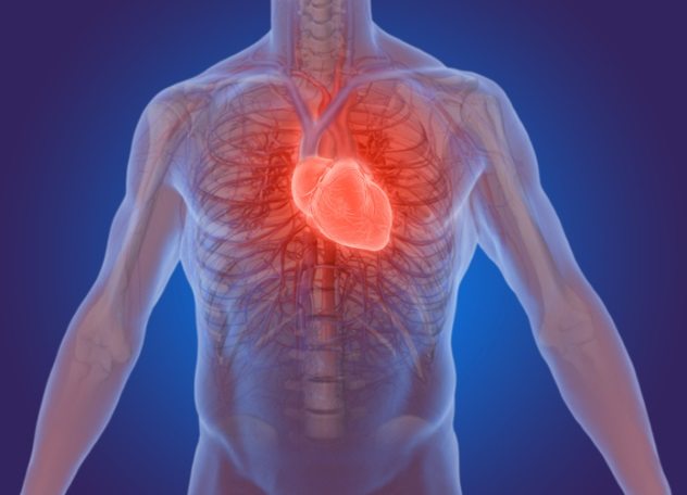
Most of us are grateful for our sphincters (or should be), but what about our valves? The cardiovascular system is essentially plumbing, and one-way valves keep things flowing in the right direction. We have four very strong pumps (the heart) which work in coordination to pump blood in a figure eight to the lungs to exchange gas and then to the rest of the body, supplying nutrients, removing waste, and keeping everything in balance.
Blood is pumped out of your heart into arteries, which expand and contract as the heart pumps. This is why you can feel a pressure wave in them, your “pulse.” As blood moves away from your heart, arteries branch into smaller and smaller vessels until they pass through extremely fine tubes called capillaries that are only a cell wide. This is when exchange happens between blood and the tissues it supplies. Blood needs to move slowly here and no longer has a pulse due to the large surface area of the microscopic capillaries.
On the way back to the heart, blood travels in veins, which converge into larger and larger vessels. However, there is not a lot of pressure driving blood back to the heart, and most of the blood needs to overcome gravity to return. To deal with this, veins have one-way valves which keep blood flowing in the right direction. Sometimes you can see valves in people’s arms, particularly when you have a tourniquet on for a blood test; they look like little knobbles along an otherwise straight vein.
There are also four essential one-way valves within the heart. Each of the four pumping chambers in the heart has a one-way valve which snaps shut when it contracts to prevent blood from being pumped out in the wrong direction. The chambers in your heart work in pairs, and it is the sound of these valves snapping shut during the pumping action that you hear as the two “lub-dub” heart sounds. If there is anything wrong with how the valves work, you can hear added heart sounds, and the pump will work less effectively.[5]
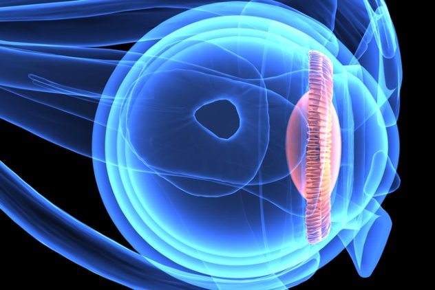
5 Lens

If you’ve ever had glasses fitted, you know how arduous the process is to find exactly the right lens to correct your vision. Much like the lenses in glasses, you have lenses within your eyes. They are transparent, concave structures that bend light to focus images onto the back of your eyeball, the retina, which sends the information to your brain to be interpreted as vision.[6]
Unlike glass or polycarbonate lenses, our anatomical lenses are elasticated and able to change their shape to focus on objects at all different distances. As we age, the lens gradually loses elasticity. This is why most people require glasses to assist with reading as they get older; the lens is less able to recoil or “bounce back” into its thickest form, which is required for near vision. Glasses help to bend the light more, prior to passing through the eye.
4 Ciliary Muscle
How exactly do our lenses manage to change shape? This is achieved by the ciliary muscle, a rim of muscle around the lens which contracts and relaxes to make the lens thicker or thinner.[7] This, in turn, bends beams of light entering the eye more or less, to keep images in focus.
This muscle movement, known as accommodation, is one of the most sophisticated motor functions in the body. Indeed, our eyes are among the most complex organs in our bodies.
3 Epiglottis

Anatomically, our trachea is in front of our esophagus, so every time we swallow, our food or water needs to pass over our windpipe and in to our food pipe. If this action is not coordinated, we choke.
The epiglottis is a flap of elastic cartilage which projects from the top of the larynx (the top part of the windpipe). When you swallow, the larynx is pulled upward. This is why you can see people’s throats move up and down when they swallow. The “Adam’s apple” is a prominence of cartilage in the larynx which makes this action more obvious in males. When the larynx is pulled upward, the epiglottis is folded over the entrance to the windpipe so that food and water pass over it, into the esophagus.
This is why it is important to lie someone on their side in the recovery position in first aid when appropriate. This is to keep their airway open and to allow any water or secretions to drain out of the mouth instead of into the airway.[8]
2 Diaphragm
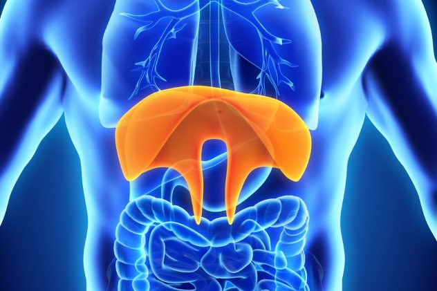
The diaphragm is a large area of fibrous and muscle tissue which separates the abdominal and thoracic cavities, and when it twitches, we get hiccups. Although the rib cage expands and contracts, diaphragm is the main muscle responsible for breathing. When relaxed, the diaphragm is dome-shaped, curving up into the thoracic cavity. When it contracts, the muscle flattens, increasing the intrathoracic volume and creating a sucking action, drawing air into the lungs as they expand.
The diaphragm also helps to regulate pressure on the chest and abdomen when vomiting, coughing, urinating, and passing stool.
When you look at an X-ray of the chest, the diaphragm is higher on the right than the left, due to the location of the liver.[9] Every time you breathe, all your abdominal contents below the diaphragm move slightly as you inhale and exhale.
1 Skin

It’s the largest organ in the body, and although it’s one of the more highly recognized body parts on the list, its importance is not. The skin has six primary roles, if any of which stopped working, you would get very sick or even die.[10]
Firstly, skin provides a barrier against physical, thermal, chemical, and radiation sources of potential trauma encountered in daily life. Also, skin regulates your body temperature. As annoying as we find sweating to be, it is actually essential to maintaining our normal physiology and is also involved in another primary function: maintaining the body’s fluid and electrolyte balance.
Skin also has multiple immune functions, acting as both a physical and immunological barrier against infection and allergic triggers. Metabolic functions of the skin include the production of vitamin D and other proteins that cells need to work. Finally, the skin is the most diverse sensory organ in the body, capable of sensing heat, cold, light and firm pressure, pain, and vibrations.
Littoral Spaces
Medicine / Arts
Read more about our surprisingly complex and amazing bodies on 10 Things We’re Just Now Learning About The Human Body and 10 Bizarre Ways The Human Body Is Used.
More Great Lists
 10 Reasons For Keeping Human Body Parts After Death
 10 Fascinating Facts And Stories Involving Body Parts
 10 Amazing Things You Didn't Know Our Body Parts Can Do
 10 Products Made From Human Body Parts And Secretions
 Top 10 Record-Breaking Body Parts
 Top 10 Body Parts We Lost To Evolution
 10 People Who Secretly Lived In Other People's Houses
 10 Governments That Secretly Have Kill Lists
 10 Famous People Who Were Secretly Spying On Us
fact checked by
Jamie Frater
More Great Lists